Amoebiasis

1. Which of the following is the causative agent of amoebiasis?
A. Giardia lamblia
B. Entamoeba histolytica
C. Balantidium coli
D. Cryptosporidium parvum
💬 Explanation: Entamoeba histolytica is the protozoan parasite responsible for amoebiasis.
2. The invasive form of Entamoeba histolytica is:
A. Precyst
B. Mature cyst
C. Trophozoite
D. Immature cyst
💬 Explanation: The trophozoite form invades colonic mucosa and causes disease.
3. The characteristic lesion of intestinal amoebiasis is:
A. Caseating granuloma
B. Flask-shaped ulcer
C. Pseudomembrane
D. Crypt abscess
💬 Explanation: Trophozoites invade the mucosa and submucosa creating flask-shaped ulcers.
4. Extraintestinal amoebiasis most commonly affects the:
A. Lungs
B. Brain
C. Liver
D. Kidney
💬 Explanation: Amoebic liver abscess is the most common form of extraintestinal amoebiasis.
5. Which of the following is the most common symptom of amoebic liver abscess?
A. Cough with sputum
B. Constipation
C. Right upper quadrant pain
D. Jaundice
💬 Explanation: RUQ pain and fever are classical features.
6. In amoebic liver abscess, the pus appears:
A. Bloody and purulent
B. Greenish and foul-smelling
C. Anchovy sauce-like
D. Clear and odorless
💬 Explanation: The pus is thick, brown, and odorless — resembling anchovy sauce.
7. Which diagnostic test is most specific for amoebic liver abscess?
A. Ultrasound
B. Serology (ELISA or IHA)
C. Stool microscopy
D. X-ray abdomen
💬 Explanation: Serological tests are highly sensitive and specific for extraintestinal amoebiasis.
8. The infective form of Entamoeba histolytica is:
A. Trophozoite
B. Pseudocyst
C. Cyst
D. Spore
💬 Explanation: Mature quadrinucleated cysts are the infective form transmitted via fecal-oral route.
9. In stool microscopy, the diagnostic finding in amoebiasis is:
A. Charcot-Leyden crystals
B. Bile-stained ova
C. Trophozoites with ingested RBCs
D. Cyst with 8 nuclei
💬 Explanation: Presence of erythrophagocytosis is diagnostic of E. histolytica.
10. Which drug is most effective against the trophozoite form in tissues?
A. Diloxanide furoate
B. Metronidazole
C. Paromomycin
D. Iodoquinol
💬 Explanation: Metronidazole is effective against tissue forms and is the drug of choice.
11. The vector responsible for transmission of E. histolytica is:
A. Anopheles mosquito
B. Tsetse fly
C. Sandfly
D. None (fecal-oral route)
💬 Explanation: Amoebiasis spreads via ingestion of cysts through contaminated food or water; it is not vector-borne.
12. Which of the following is NOT a common complication of amoebic liver abscess?
A. Rupture into peritoneum
B. Pleuropulmonary involvement
C. Acute pancreatitis
D. Rupture into pericardium
💬 Explanation: Pancreatitis is unrelated to amoebiasis; other options are well-known complications.
13. Best imaging modality to diagnose amoebic liver abscess:
A. Chest X-ray
B. Ultrasound abdomen
C. CT brain
D. Colonoscopy
💬 Explanation: Abdominal ultrasound is the most common, cost-effective, and sensitive method for detecting liver abscesses.
14. Stool microscopy in amoebiasis shows cysts with how many nuclei?
A. 2
B. 3
C. 4
D. 6
💬 Explanation: Mature infective cysts of E. histolytica are quadrinucleated (4 nuclei).
15. Which of the following is used to eradicate intestinal cysts of E. histolytica?
A. Diloxanide furoate
B. Metronidazole
C. Tinidazole
D. Ornidazole
💬 Explanation: Diloxanide furoate is a luminal amoebicide used after metronidazole to eradicate intestinal cysts.
16. Which stain is commonly used for identifying E. histolytica cysts in stool?
A. Ziehl-Neelsen stain
B. Gram stain
C. Trichrome stain
D. Silver stain
💬 Explanation: Trichrome staining enhances visualization of trophozoites and cysts of E. histolytica under light microscopy.
17. Which of the following is a distinguishing feature of E. histolytica trophozoites?
A. Pseudohyphae
B. Ingested red blood cells (erythrophagocytosis)
C. Sulfur granules
D. Maltese cross
💬 Explanation: Erythrophagocytosis is a pathognomonic feature of E. histolytica, seen in its trophozoite form.
18. What is the most common site for extraintestinal amoebiasis?
A. Brain
B. Liver
C. Spleen
D. Lungs
💬 Explanation: Liver is the most frequent site of extraintestinal amoebiasis, commonly forming abscesses.
19. Which of the following conditions mimics amoebic colitis the most?
A. Pseudomembranous colitis
B. Crohn’s disease
C. Ulcerative colitis
D. Ischemic colitis
💬 Explanation: Amoebic colitis closely mimics ulcerative colitis both clinically and endoscopically.
20. E. histolytica trophozoites are best identified in:
A. Formed stool sample
B. Freshly passed liquid stool
C. Rectal biopsy
D. Sputum
💬 Explanation: Trophozoites are labile and rapidly degenerate; they are best seen in fresh, liquid stools within 30 minutes of collection.

20-Point Summary Table: Amoebiasis

🔢 No.🧠 Topic📌 Key Point
1️⃣DefinitionAmoebiasis is an intestinal infection caused by Entamoeba histolytica.
2️⃣Causative AgentEntamoeba histolytica, a pathogenic protozoan amoeba.
3️⃣ReservoirInfected humans (asymptomatic cyst passers and patients).
4️⃣Mode of TransmissionFeco-oral route via contaminated food/water or hands.
5️⃣Infective FormMature quadrinucleate cyst of E. histolytica.
6️⃣Site of InfectionPrimarily the colon (cecum, ascending colon, rectum).
7️⃣Trophozoite FeaturesMotile, with single nucleus, ingests RBCs (pathognomonic).
8️⃣Cyst FeaturesNon-motile, 1–4 nuclei, infective in nature.
9️⃣Asymptomatic CarrierCan transmit infection via cysts in stool.
🔟Intestinal AmoebiasisPresents as diarrhea, abdominal pain, and dysentery.
1️⃣1️⃣Amoebic DysenteryBloody mucoid stools, tenesmus, cramping — no fever usually.
1️⃣2️⃣Extraintestinal DiseaseAmoebic liver abscess is the most common complication.
1️⃣3️⃣Liver Abscess FeaturesRight lobe > left, tender hepatomegaly, “anchovy sauce” pus.
1️⃣4️⃣Diagnosis (Stool)Microscopy shows trophozoites with ingested RBCs.
1️⃣5️⃣Diagnosis (Serology)Anti-amoebic antibody tests (useful in liver abscess).
1️⃣6️⃣Imaging (Liver Abscess)Ultrasound or CT reveals hypoechoic lesion in liver.
1️⃣7️⃣Medical TreatmentMetronidazole + luminal agent (diloxanide furoate).
1️⃣8️⃣Surgical IndicationsRare — done for rupture of abscess into peritoneum.
1️⃣9️⃣PreventionSafe water, hand hygiene, sanitation, avoid raw salads.
2️⃣0️⃣Important DifferentialsBacillary dysentery, IBD, other parasitic infections.

1. What is Amoebiasis?
• Amoebiasis is caused by Entamoeba histolytica, a protozoan parasite.
• It mainly affects the colon and causes dysentery or diarrhea.
• Transmission is through ingestion of cysts in contaminated food or water.
• It may be asymptomatic or progress to severe disease.
• Extraintestinal spread can lead to liver abscess.

2. How is Entamoeba histolytica transmitted?
• Fecal-oral route via contaminated water or food.
• Cysts survive outside the host and are infective.
• Poor sanitation and hygiene promote transmission.
• Flies can mechanically spread cysts to food.
• Person-to-person spread may occur in crowded areas.

3. What are the main clinical features of intestinal amoebiasis?
• Symptoms include diarrhea, abdominal cramps, and tenesmus.
• Stools may contain blood and mucus.
• Weight loss and fatigue in chronic cases.
• Severe colitis may resemble IBD.
• Perforation and toxic megacolon are rare complications.

4. What is amoebic liver abscess?
• A liver abscess caused by invasive E. histolytica.
• Presents with fever, right upper abdominal pain, hepatomegaly.
• Often solitary and in the right lobe of liver.
• Anchovy sauce pus on aspiration.
• May rupture into pleura or peritoneum if untreated.

5. How is amoebiasis diagnosed?
• Stool microscopy for cysts/trophozoites.
• Antigen detection tests and PCR improve accuracy.
• Serology helps in extraintestinal disease.
• Ultrasound/CT for liver abscess evaluation.
• Biopsy in cases mimicking colitis.

6. What are the forms of E. histolytica seen in humans?
• Two forms: cyst (infective) and trophozoite (invasive).
• Cysts are passed in stool and survive outside.
• Trophozoites found in tissue and cause damage.
• Cysts have 1–4 nuclei; trophozoites have a central karyosome.
• Both are essential in transmission and pathology.

7. What is flask-shaped ulcer in amoebiasis?
• A classical histological lesion in colonic amoebiasis.
• Ulcer has narrow neck and broad base.
• Caused by trophozoite invasion and necrosis.
• Seen in large intestine on biopsy.
• Helps differentiate from bacterial ulcers.

Flask-shaped ulcers are a characteristic pathological feature of invasive amoebiasis, particularly amoebic colitis, caused by the parasite Entamoeba histolytica

Here’s a breakdown of how they form and their significance:

Formation

  1. Invasion: Pathogenic E. histolytica trophozoites, released from cysts in the intestines, invade the crypts of the colonic glands and burrow into the submucosa.
  2. Lateral Extension: The invasion is typically halted by the muscularis propria, causing the trophozoites to spread laterally within the submucosa.
  3. Inflammation and Necrosis: This lateral burrowing and the host’s inflammatory response lead to necrosis and the development of the characteristic flask-shaped ulcer.
  4. Undermining the Mucosa: The base of the ulcer is broader than the apex, undermining the overlying mucosa. 

Key features

  • Shape: Resembles a flask or bottle with a narrow neck and a broader base.
  • Location: Commonly found in the cecum and ascending colon, but can occur anywhere in the colon.
  • Histology: Trophozoites are often seen at the base of the ulcer and within the submucosa, along with necrotic debris and inflammatory cells. 

Significance in diagnosis

  • The presence of flask-shaped ulcers on colonoscopy and/or biopsy is highly suggestive of amoebic colitis.
  • However, it’s important to note that other conditions like Balantidium coli (Balantioides coli) infection can also cause similar ulcers.
  • Therefore, definitive diagnosis relies on identifying E. histolytica trophozoites in tissue biopsies, stool samples, or through molecular tests like PCR. 

Differentiation from other conditions

  • Amoebic colitis can sometimes resemble other conditions like inflammatory bowel disease (IBD).
  • Features that can help differentiate amoebiasis from IBD include:
    • Discrete ulcers with intervening normal mucosa in amoebiasis, while IBD often presents with continuous inflammation.
    • The presence of necrotic material and trophozoites lining the ulcers in amoebiasis.
    • Specific identification of E. histolytica through appropriate testing. 

In conclusion, the flask-shaped ulcer is a distinctive feature of invasive amoebiasis, offering crucial clues for diagnosis when observed through endoscopic examination and confirmed by histopathological analysis or specific laboratory tests. 
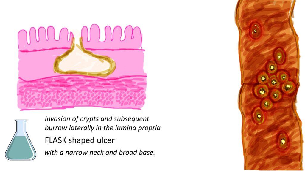


**Amoebiasis** is caused by trophozoite invasion and necrosis, typically seen in the large intestine on biopsy. This helps differentiate it from bacterial ulcers. Amoebiasis results from trophozoite invasion and necrosis in the large intestine on biopsy. This helps distinguish it from bacterial ulcers. The flask-shaped ulcers are a characteristic feature of invasive amoebiasis caused by Entamoeba histolytica. They offer essential clues for diagnosis during endoscopic examination. Confirmation is done through histopathological analysis or specific laboratory tests.